Embryo Development 101
April 30, 2010
When an egg is retrieved from the ovaries, it is fertilized either through putting the egg and sperms in the same space (usually a petri dish) so it will naturally fertilized or through a process called ICSI, where one sperm is injected into an egg. Once the egg and sperm had a chance to merge, the new product is called an embryo.
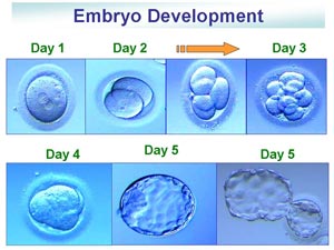
Here is a pictorial view and explanation of what our embryos are expected to look like day to day and the names used (by the way — this is the same whether it is inside the body or outside the body):
Post egg retrieval
Day 1: 1 cell with 2 dots in the middle. This indicates a normally fertilized egg with each dot representing genetic materials from the mom and the dad.
Day 2: usually 2-4 cells. The cells are expected to double every 24 hours or so.
Day 3: usually 6-8 cells. Sometimes more cells. Same doubling effect. This is when fragmentation can be observed (small dots, not clean cell divisions) representing abnormal cells. I’ve read somewhere that chromosonal abnormality can be around 25-70%.
Day 4: Morula or compacting cell stage. This is when the cells have doubled to around 16 and the individual balls are starting to expand and blend with the others. You can see the bumps still.
Day 5: Blastocyst. This is when we see 3 distinct parts – the shell, one dense bunch of cells (the baby), and the lighter cells (the placenta). The blastocyst is ready for implantation but first it has to hatch from the shell. The picture on the bottom right is a hatching blastocyst (can happen naturally or with the help of your embryologist – assisted hatching, like ours in IVF#1). The hatched blastocyst should implant within 12-24 hours to the uterine lining.
